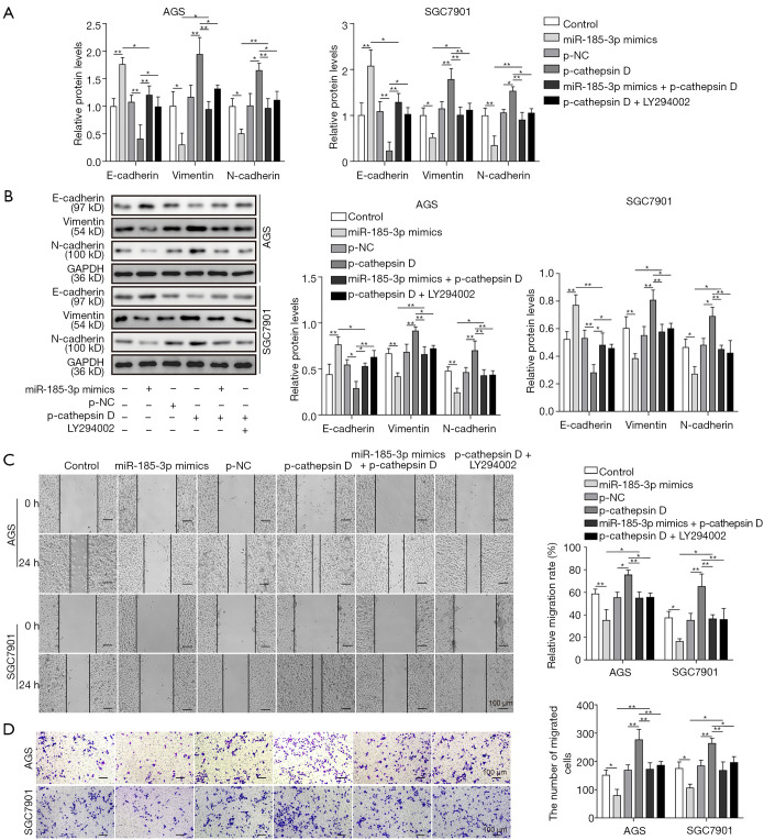
Figure 5.
MiR-185-3p regulated EMT via cathepsin D mediated PI3K/Akt signaling pathway. (A) qRT-PCR analysis of EMT markers in AGS and SGC7901 cells after transfection with miR-185-3p mimics, p-cathepsin D, miR-185-3p mimics and p-cathepsin D, as well as p-cathepsin D followed by administration of LY294002. (B) Western blotting of EMT markers in AGS and SGC7901 cells after transfection. The quantification of EMT markers was normalized against GAPDH. (C) Cell scratch assay of AGS cells after transfection. Left, images of the migration of cells after incubation for 24 h; right, quantification of the cell migration. (D) Transwell assay of AGS cells after transfection. Left, images of migration cells in the lower chamber; right, cell counts of the migration cells in the lower chamber. *, P<0.05; **, P<0.01.

