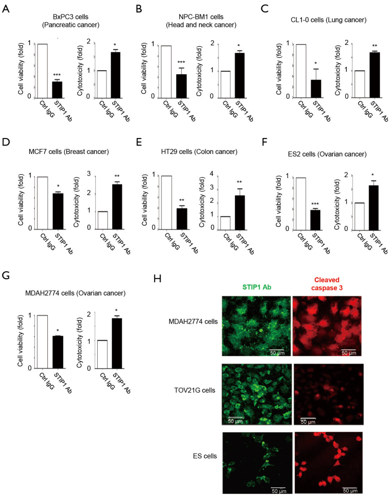
Figure 2.
HEPES-mediated intracellular delivery of anti-stress-induced phosphoprotein 1 (STIP1) antibodies inhibits viability and increases toxicity in different cancer cell lines. Control IgG2 antibodies (Ctrl IgG) or anti- STIP1 antibodies (STIP1 Ab) (Abnova; Cat. No: H00010963-M02) were initially mixed with appropriate concentrations of HEPES (reported in parentheses) 15 min before intracellular delivery. Antibodies were subsequently delivered into (A) pancreatic cancer BxPC3 (30 mM HEPES), (B) head and neck cancer NPC-BM1 (20 mM HEPES), (C) lung cancer CL1-0 (20 mM HEPES), (D) breast cancer MCF7 (20 mM HEPES), (E) colon cancer HT29 (20 mM HEPES), (F) ovarian cancer ES2 (20 mM HEPES), and (G) ovarian cancer MDAH2774 (20 mM HEPES) cells. After 24−48 h of incubation, cell viability (24 h) and cytotoxicity (48 h) were analyzed with the [3-(4,5-Dimethylthiazol-2-yl)-2,5-diphenyltetrazolium bromide] (MTT) and lactate dehydrogenase (LDH) assays, respectively. (H) The detection of anti-STIP1 antibodies in MDAH2774, TOV21G and ES2 cells were performed by staining with Alexa Fluor 488 anti-mouse IgG antibodies, whereas cleaved caspase 3 antibodies were imaged using Alexa Fluor 546 anti-rabbit IgG antibodies. Scale bars indicate 25 µm. Error bars indicate the standard errors of the mean (n=3). *P<0.05, **P<0.01, and ***P<0.001.

