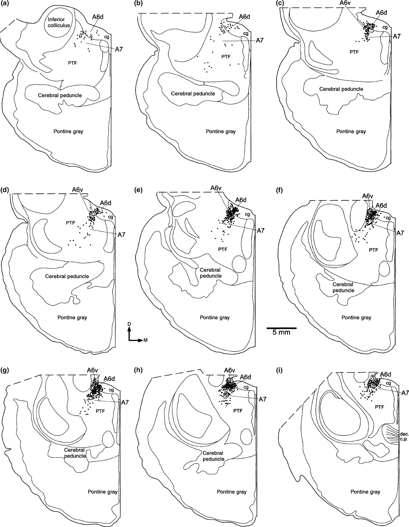
Figure 1.

Diagrams of the cellular distribution of the tyrosine hydroxylase immunoreactive cells which make up the locus coeruleus complex of the bottlenose dolphin. Sections are approximately 1 mm apart and are shown from rostral (a) to caudal (i). Each dot represents a single immunoreactive cell. cg, central, or peri-aqueductal gray matter; D, dorsal; dec c.p., decussation of the cerebral peduncle or brachium conjunctivum; PTF, pontine tegemental field.
