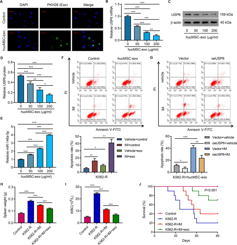
Fig. 6. HucMSC exosome promoted IM-induced apoptosis in K562-R cells.
A PKH26-labeled hucMSC-exo was uptaken by K562-R cells. Scale bar: 50 μm. B–E The expression of USP6 and miR-146a-5p in K562-R cells treated by different hucMSC-exo concentrations (0, 50, 100, and 200 μg/mL) for 24 h. F Flow cytometry analysis of apoptosis of K562-R cells treated by hucMSC-exo (100 μg/mL) alone or in combination with 1 μM IM for 48 h. G Flow cytometry analysis of apoptosis of USP6-overexpressing K562-R cells treated by hucMSC-exo (100 μg/mL) alone or in combination with 1 μM IM for 48 h. In vivo tumor formation assays. K562-R was administered to each mouse. On day 90, mice were sacrificed and (H) spleen weight, (I) white-blood cell count, and (J) survival curve were analyzed. *P < 0.05, **P < 0.01, ***P < 0.001.

