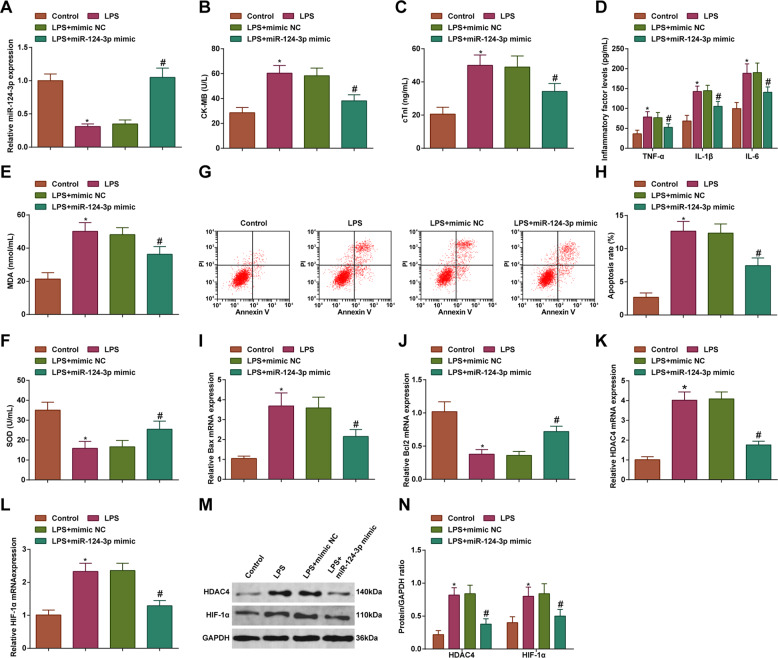
Fig. 3. Upregulation of miR-124-3p ameliorates inflammation, oxidative stress, and apoptosis of LPS-treated cardiomyocytes.
A RT-qPCR detected miR-124-3p expression level in cardiomyocytes; B ELISA detected CK-MB level in cardiomyocytes; C ELISA detected cTn-I level in cardiomyocytes; D ELISA detected TNF-α, IL-1β, and IL-6 levels in cardiomyocytes; E MDA level in cardiomyocytes; F SOD activity in cardiomyocytes; G and H Flow cytometry detected apoptosis of cardiomyocytes; I RT-qPCR detected Bax mRNA expression level in cardiomyocytes; J RT-qPCR detected Bcl-2 mRNA expression level in cardiomyocytes; K RT-qPCR detected HDAC4 mRNA expression level in cardiomyocytes; L RT-qPCR detected HIF-1α mRNA expression level in cardiomyocytes; M and N Western blot detected HDAC4 and HIF-1α protein expression in cardiomyocytes; the measurement data were expressed as mean ± standard deviation, repetition = 3, *P < 0.05 compared with the control group; #P < 0.05 compared with the LPS + mimic NC group.

