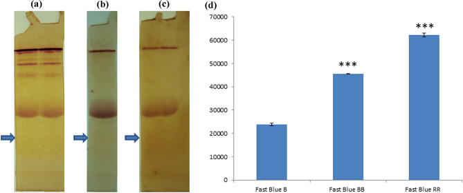
Fig. 1.
Representative picture of native PAGE of plasma proteins followed by staining with 2NA for detection of esterase activity. The gel is incubated with 75 µM 2NA in the solution of a Fast Blue B (0.12%), b Fast Blue BB (0.12%), c Fast Blue RR (0.12%) for overnight staining respectively. The densitometry analysis of the background of the gel (→). Mean ± SD (n = 6) of the background density is shown in d for (A), (B) and (C) respectively (Y-axis in square pixel where 96 pixels = 1 inch). ***p value less than 0.0001

