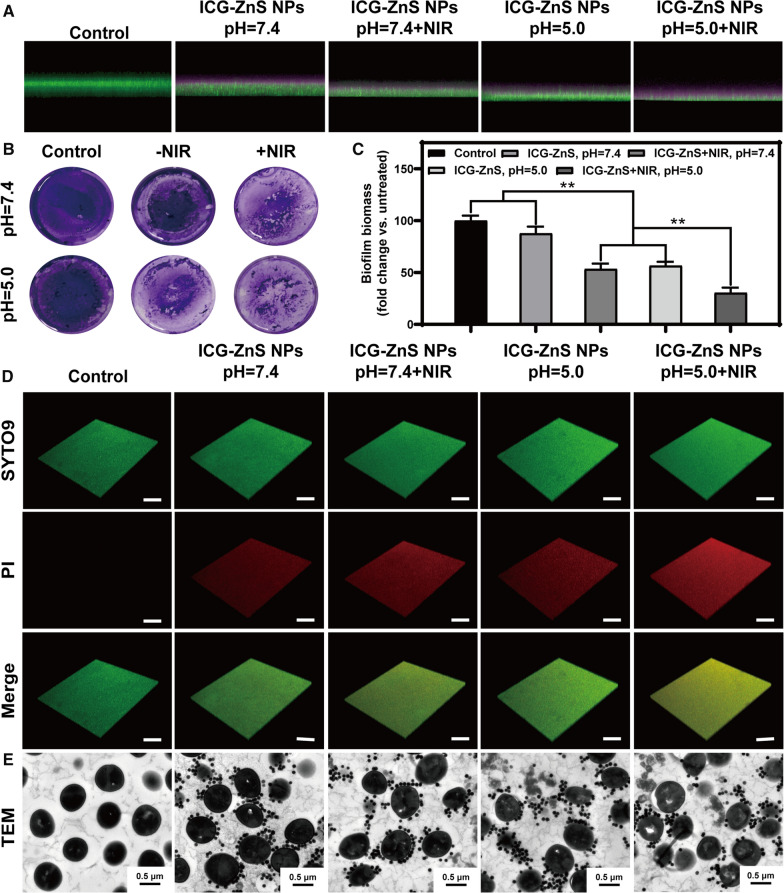
Fig. 5.
In vitro penetration assay anti-biofilm activity toward MRSA biofilms. A CLSM images of MRSA biofilms after treated with ICG-ZnS NPs and ICG-ZnS NPs + NIR at different concentrations at pH 7.4 and 5.0. The presence of biofilm was detected with SYTO 9 dye (green channel), and the nanoparticles were detected with ICG (pink channel). B Crystal violet assay to assess the anti-biofilm activity of the samples against an MRSA biofilm. C Quantitative analysis of the crystal violet-stained biofilms in panel a by measuring the absorbance at 590 nm. D Live/dead staining images of MRSA biofilm using 3D CLSM (scale bar: 100 μm). E Typical TEM images of MRSA biofilm treated with different groups (scale bar: 0.5 μm). Data are presented as the means ± SD (n = 3), *P < 0.05, **P < 0.01

