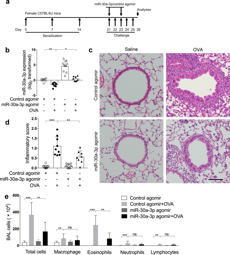
Fig. 6.
MiR-30a-3p overexpression suppresses airway eosinophilic inflammation in a mouse model of allergic airway inflammation. a Experimental schedule. mmu‐miR‐30a‐3p or control agomir was administered intranasally 2 h before OVA challenge on days 21 and 23. b The transcript levels of miR-30a-3p in mouse lungs were determined by quantitative PCR. The transcript levels were expressed as log2 transformed and relative to the mean of control group. c Representative H&E staining of mouse lung sections. Scale bar, 50 μm. d Lung inflammatory scores were calculated as described in ‘Materials and methods’. e Counts for macrophages, eosinophils, lymphocytes and neutrophils in BAL fluid. n = 7–10 mice per group. Data are mean ± SD. *P < 0.05; **P < 0.01; ***P < 0.001 (one-way ANOVA followed by Tukey’s multiple comparison test)

