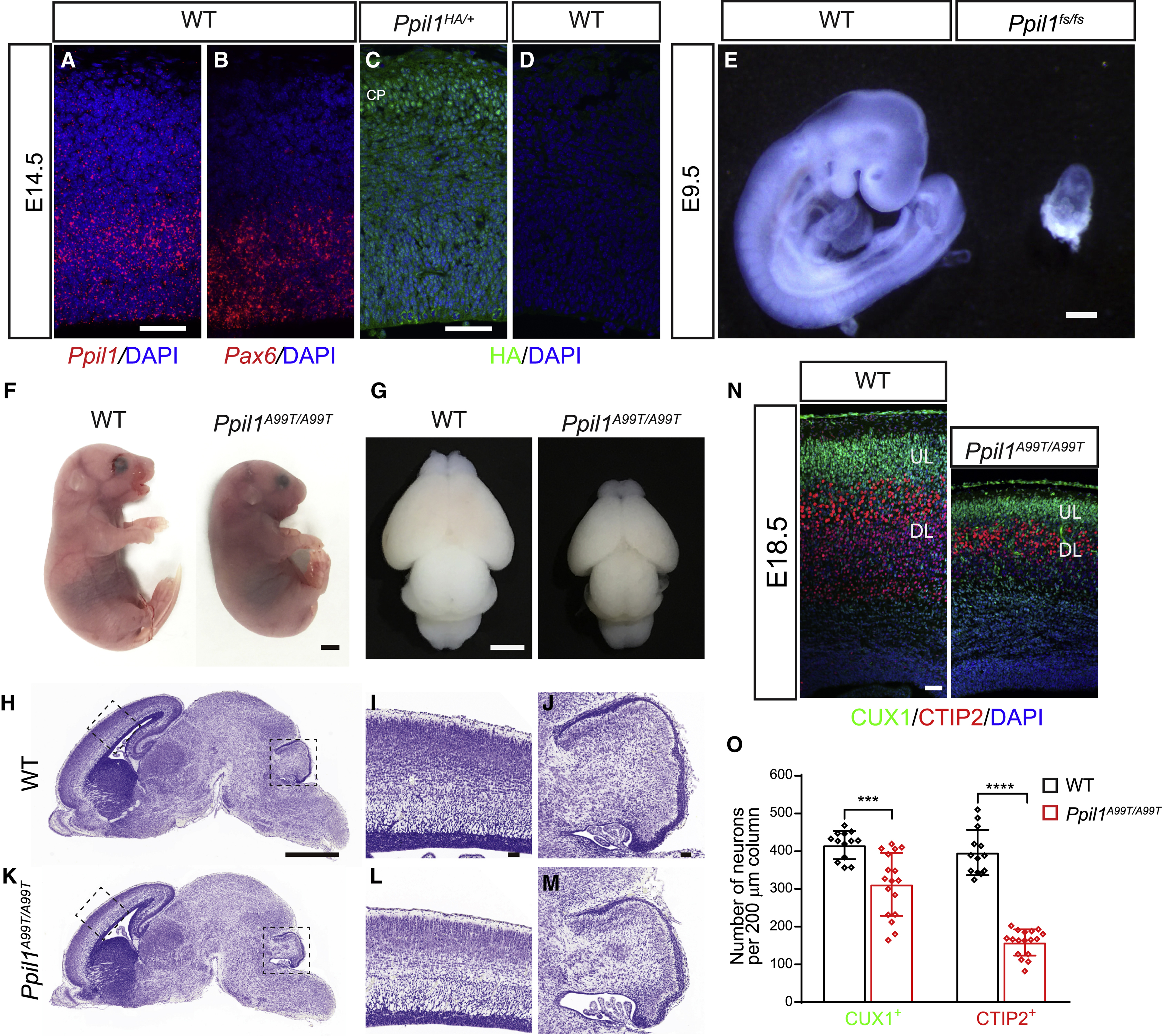
Figure 2. Patient PPIL1 mutation knockin mice exhibit PCHM-like phenotype.

(A and B) Fluorescent in situ hybridization (FISH) on coronal sections of E14.5 brain cortex hybridized with Ppil1 (A) and Pax6 (B) probes using RNAscope. Scale bar: 50 μm.
(C and D) Coronal sections of E14.5 embryos from Ppil1HA/+ (C) and WT (D) embryos immunostained with an anti-HA antibody showing ubiquitous expression of PPIL1. CP: cortical plate. Bar: 50 μm.
(E) Ppil1 fs/fs mouse embryos showed reabsorption at E9.5. Bar: 2 mm.
(F and G) Homozygous patient variant p.A99T knockin mouse with microcephaly at E18.5.
(H-M) Nissl stained sagittal sections of E18.5 Ppil1A99T/A99T brains, magnified for dashed regions in the cerebral cortex and cerebellum.
(N) E18.5 Ppil1A99T/A99T cortex (coronal) shows reduced thickness but with intact lamination based upon immunostaining against CUX1 (upper layer neurons) and CTIP2 (lower layer neurons).
(O) Reduced density of cortical CUX1+ and CTIP2+ neurons in E18.5 Ppil1A99T/A99T cortex. n = 4 mice/genotype. Mean ± s.d.; p = 0.0003 CUX1+ cells; p < 0.0001 CTIP2+ cells; two-tailed unpaired t-test.
Scale bar: 1 mm in H and K; 50 μm in I, J, and L–M.
See also Figure S2.
