Figure 3.
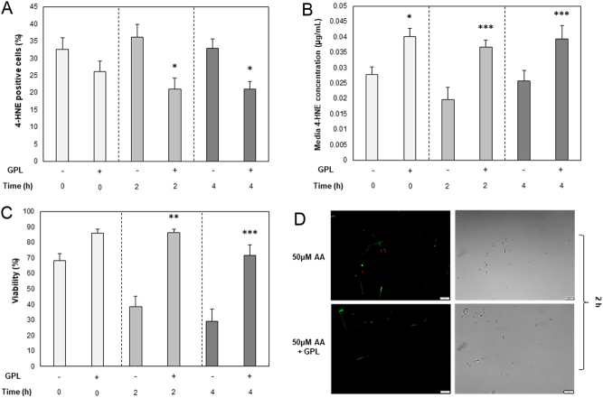
Analysis of 4-HNE adduction. (A) Total cellular 4-HNE adduction, utilizing α-HNE primary and Alexa Fluor 488 secondary antibodies, (B) Concentration of 4-HNE adducts within the spent sperm storage media and, (C) Total cell viability measured with LIVE/DEAD™ fixable far red dead cell stain obtained after co-incubation in the presence of 50 µM arachidonic acid only (AA; −) or in the presence of 50 µM AA with the addition of GPL (+), measured at regular 2 h intervals over a total of 4 h. (D) Fluorescent and phase images of spermatozoa depicting 4-HNE adduction (green) utilizing α-HNE primary and Alexa Fluor 488 secondary antibodies, after 2 h incubation with 25 µM AA only (top) and 25 µM AA with the addition of GPL (Bottom). Red fluorescence (PI) indicates a dead cell. Original magnification ×400. All data are displayed as means ± s.e.m. (*P ≤ 0.05, **P ≤ 0.01, ***P ≤ 0.001; Student’s t-test).

 This work is licensed under a