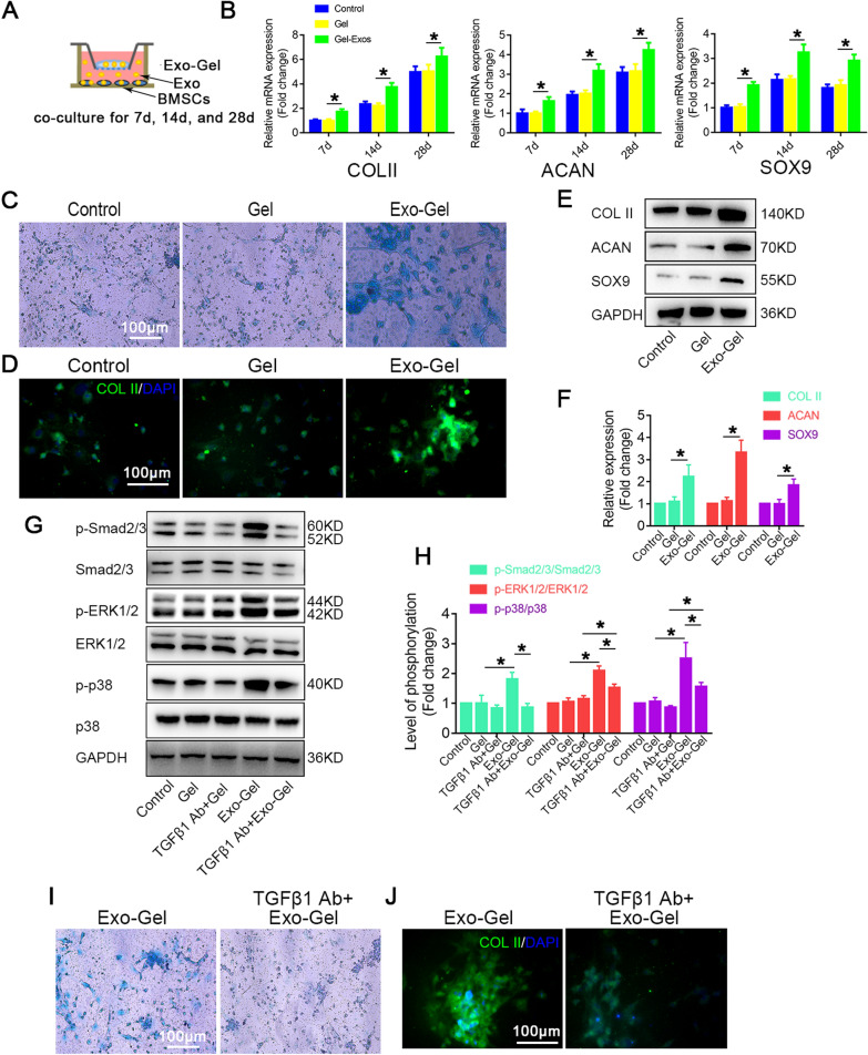
Fig. 3.
Exo-Gel enhanced chondrogenic differentiation mBMSCs in vitro. A In vitro estimate the ability of Exo-Gel by coculture in chondrogenic differentiation of mBMSCs. B RT-PCR analysis for COL II, ACAN and SOX9 mRNA in mBMSCs treated as described in (A) at different time-points. Data are represented as means ± SEM (n = 3). C AB staining of mBMSCs in different treatment groups under light microscopy on the 14th day of induction of differentiation (bar = 100 μm). D Immunofluorescent assay of mBMSCs for COL II (red) and DAPI (Blue) in different treatment groups at the same time-point in C (bar = 100 μm). E Western blot assay to detect the protein expression levels of COL II, ACAN and SOX9 at the same time-point in C. F Quantification of protein expression in E. GAPDH was used as loading control. Data are expressed as means ± SEM (n = 3). G Representative Western blot showing the levels of total protein and phosphorylated protein for the indicated molecules (Smad2/3, ERK1/2, and p38) in mBMSCs with or without TGFβ1 neutralizing antibody at the same time-point in C. H Quantification of protein expression and the level of signaling activation in G. GAPDH was used as loading control. Data are expressed as means ± SEM (n = 3). I AB staining of mBMSCs with or without TGFβ1 neutralizing antibody under light microscopy on the 14th day of differentiation (bar = 100 μm). J Immunofluorescent assay of mBMSCs for COL II (red) and DAPI (blue) under the same condition as (I) (bar = 100 μm). *p < 0.05

