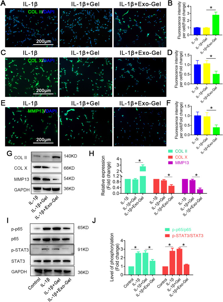
Fig. 6.
Exo-Gel suppressed IL-1β-triggered chondrocyte degeneration and enhanced anabolism in vitro. A Immunofluorescent assay to detect the expression of COL II (Gree), COL X (Green), and MMP13 (Green) in chondrocytes in different treatment groups, DAPI (blue) staining for cell nuclei (bar = 200 μm). B Quantification of COL II, COL X, and MMP13 fluorescence intensity in A. Data are expressed as means ± SEM (n = 6). C Representative Western blots of COL II, COL X, and MMP13 expression in chondrocytes in different groups. D Quantification of COL II, COL X, and MMP13 protein expression in chondrocytes in C. GAPDH was used as loading control. Data are expressed as means ± SEM (n = 3). E Representative Western blot showing the levels of total protein and phosphorylated protein for the indicated molecules (p65 and STAT3) in chondrocytes in different treatment groups. F Quantification of protein expression and the level of signaling activation in E. GAPDH was used as loading control. Data are expressed as means ± SEM (n = 3) *p < 0.05

