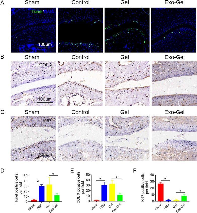
Fig. 9.
Exo-Gel inhibited cartilage degeneration of subtalar joint. A TUNEL (Green) staining in the subtalar joint section to analyze chondrocyte apoptosis at 4 weeks postoperatively, DAPI (blue) staining for cell nuclei (bar = 100 μm). B, C immumohistochemical staining of COL X (B) and Ki67 (C) in the subtalar joint section (bar = 100 μm). (D) Quantitative assessment of number of apoptotic cells in A. Data are expressed as means ± SEM (n = 4). (E, F) quantitative analysis of COL X and Ki67 positive cells in cartilage tissue in B and C, respectively. Data are expressed as means ± SEM (n = 4). *p < 0.05

