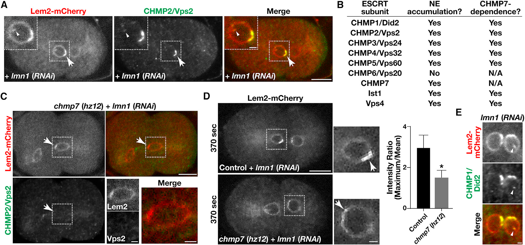
Figure 6. ESCRT-III accumulates on INM invaginations.

(A, C, and E) Embryos expressing a mCherry fusion to LEM2 (red) were fixed and stained using antibodies directed against CHMP2/Vps2 (green; A and C) or CHMP1/Did2 (green; E) and imaged using confocal microscopy following partial depletion of lamin in the presence (A and E) or absence (C) of CHMP7. Images are representative of more than 10 embryos analyzed. Bars, 10 μm (A and C) or 2 μm (E); inset bars, 2 μm.
(B) A summary highlighting the ESCRT-III components that accumulate on the NE together with LEM2 and their dependence on CHMP7.
(D) Embryos expressing a mCherry fusion to LEM2 were imaged live using confocal microscopy 370 s after anaphase onset, following partial depletion of lamin in the presence and absence of CHMP7. Images are representative of more than 10 embryos analyzed. Bar, 10 μm; inset bar, 2 μm. Hyperaccumulation of LEM2 on NE subdomains was quantified under both conditions (right). Error bars represent mean ± SEM. *p < 0.05, as compared with control, using a t test.
See also Figure S3.
