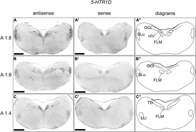
FIGURE 5.
In situ hybridization of 5-HTR1D in the P1 chick brainstems. Digoxigenin-labeled RNA antisense (A–C) and sense (A’–C’) 5-HTR1D probes were used for in situ hybridization in coronal sections of P1 chick brains. To evaluate the expression patterns of 5-HTR1D, sections of five chicks were analyzed, and representative images of two chick brain sections are shown. (A”–C”) Diagrams of coronal sections are shown in the rightmost panels. The levels of the sections (A1.8, A1.6, and A1.4) are in accordance with those mentioned in Kuenzel and Masson’s chick atlas (Kuenzel and Masson, 1988). FLM, fasciculus longitudinalis medialis; GCt, griseum centrale; LLi, nucleus ventralis lemnisci lateralis; nIV, nucleus nervi trochlearis; SLu, nucleus semilunaris; TD, nucleus tegmenti dorsalis; P1, post-hatch day 1. Scale bars = 1 mm.

