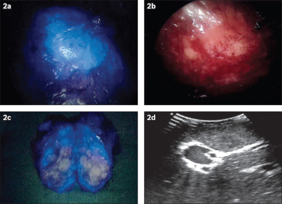
Fig. 2.

A hepatocellular carcinoma (HCC) located in segment 6 during open segment 6 wedge resection. Intraoperative (a) fusion fluorescence image and (b) NIR camera photograph show (a & b) the HCC visualised intraoperatively following a preoperative intravenous injection of indocyanine green. (c) Fluorescence image shows the resected specimen with a rim-enhancing pattern. (d) US image shows the HCC lesion intraoperatively.
