
Fig. 11.
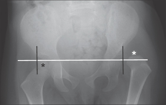
Pelvic radiograph shows developmental dysplasia of the hip on the left where the femoral epiphysis (white asterisk) lies in the superolateral quadrant (lateral to Perkin’s line, black; and above Hilgenreiner’s line, white). It should normally be in the inferomedial quadrant (black asterisk).