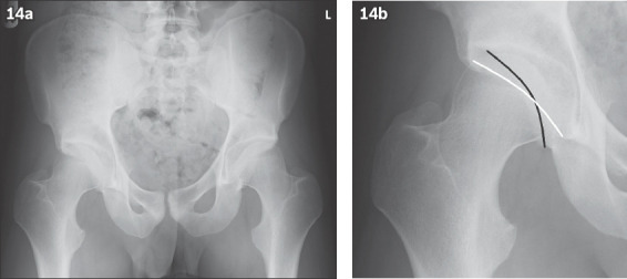
Fig. 14.

(a & b) Pelvic radiographs show an example of bilateral acetabular retroversion. The crossover sign is seen in (b), where the posterior wall (black line) of the acetabulum is seen to cross over and project medial to the anterior wall (white line) of the acetabulum.
