
Fig. 17.
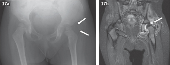
A child with left hip joint effusion. (a) Pelvic radiograph shows the gluteal fat stripe bulging superiorly (arrows) due to the hip joint effusion. (b) Coronal MR short tau inversion recovery image of the pelvis shows the presence of the left hip effusion (arrow).