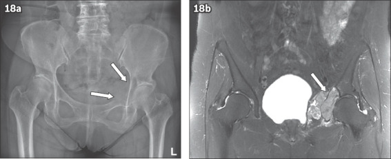
Fig. 18.

(a) Pelvic radiograph shows a destructive lytic bone lesion (arrows) in the left acetabulum with discontinuity of the iliopectineal line. (b) Subsequent coronal MR short tau inversion recovery image of the pelvis shows soft tissue metastatic infiltrate causing bony destruction of the left acetabulum (arrow).
