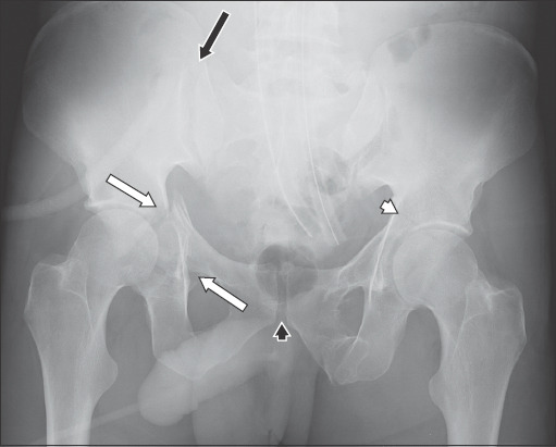
Fig. 4.

Pelvic radiograph shows complex pelvic fractures with posterior and anterior columns fractures of the right acetabulum, and disruption of the right ilioischial and iliopectineal lines (white arrows); and anterior left acetabular column fracture with discontinuity of the left iliopectineal line (white arrowhead). There is widening of the right sacroiliac joint (black arrow) and minor widening of the pubic symphysis (black arrowhead).
