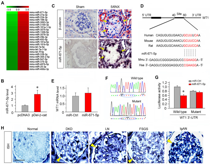
FIGURE 1.
miR-671-5p is induced by β-catenin in podocytes and targets WT1. (A) Microarray chip analysis of miRNAs expression in different groups (left: pcDNA3 control group; right: pDel-β-cat group). The red and green colors indicated high or low expression, respectively. (B) Mouse podocytes (MPC5) were transfected with empty vector (pcDNA3) or β-catenin expression plasmid (pDel-β-cat) for 24 h. qRT-PCR analysis showed the expression of miR-671-5p in different groups. *p < 0.05 (n = 3). (C) Co-localization of β-catenin and miR-671-5p in glomerular podocytes of diseased kidney. Kidney serial sections (3 μm) of 5/6NX mice were subjected to immunostaining for β-catenin and in situ hybridization for miR-671-5p. Representative micrographs from sham group are also shown. Arrows indicate positive staining in podocytes. Scale bar, 20 µm. (D) Bioinformatics analysis shows the predicted binding sites of miR-671-5p in the WT1 3′-untranslatd region (UTR) using the TargetScan software. (E) qRT-PCR analysis shows that overexpression of miR-671-5p did not affect WT1 mRNA level in mouse podocytes. MPC5 cells were transfected with miRNA negative control (miR-Ctrl) or miR-671-5p mimics (miR-671-5p) for 24 h. (F) Sequence validation of the wild type or mutant WT1 3′-UTR for the luciferase reporter construction. The wild-type miR-671-5p binding site in WT1 3′-UTR (upper) and the mutated one (bottom) in the region corresponding to the miR-671-5p seed sequence are shown. (G) Luciferase reporter assay show that miR-671-5p mimics decreased the luciferase activity in 293T cells co-transfected with wild-type WT1 3′ UTR, but not with mutant WT1 3′ UTR. *p < 0.05. (H) Representative micrographs show miR-671-5p expression in glomerular podocytes of human kidney biopsies from the patients with various CKDs by in situ hybridization. Arrowheads indicate the positive staining for miR-671-5p in glomerular podocytes. Kidney tissues adjacent to renal cell carcinoma from patients who underwent carcinoma resection were used as normal control. Scale bar, 20 µm.

