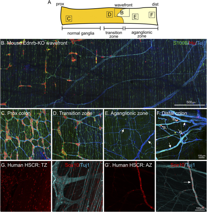
FIGURE 5.
Enteric glia are present in the gut in the absence of enteric neurons: the aganglionic zone of Hirschsprung Disease (HSCR) tissue. Whole-mount immunohistochemistry of Ednrb-KO mouse (A–F) and human HSCR patient tissue (H-H′). (A): Diagram of mouse colon including the ENS wavefront and showing the regions with “normal” ganglia, the transition zone (TZ), and the aganglionic zone (AZ). (B–F): Representative images from each of these regions, following immunohistochemistry against S100B (green), HuC/D (red) and neuronal class III β−tubulin (Tuj1, blue). The majority of S100B + glia in the aganglionic region follow nerve bundles, both large and small (arrows). Sometimes, individual Hu + neurons are present in the aganglionic zone (open arrows). NB. These are not “skip segments” as there are no clear ganglia, instead, they are just a handful of individual neuronal cell bodies. (G-G’): Patient HSCR samples labelled against Sox10 (red) and Tuj1 (blue). In the aganglionic zone, Sox10 + glia are also associated with nerve processes. The same proximal—distal orientation has been maintained in all tissues.

