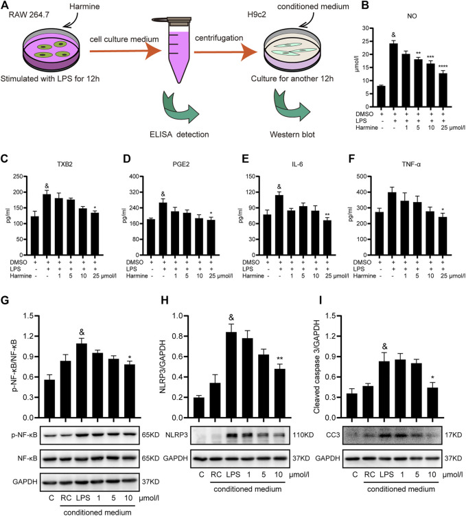
FIGURE 2.
Harmine attenuated inflammation in RAW 264.7 cells and conditioned medium-treated H9c2 cells. (A) Diagram of conditioned medium treatment of H9c2 cells. RAW 264.7 cells were pretreated with harmine (5 and 25 μmol/L) for 1 h and then stimulated with LPS (1 μg/ml) for 12 h. The medium was collected in EP tubes and then centrifuged. The supernatants were subjected to ELISAs, and harmine-conditioned medium (1, 5, or 10 μmol/L) was subsequently incubated with H9c2 cells for another 12 h. H9c2 cell lysates were collected for western blotting after culture. (B–F) The levels of the following inflammatory mediators in the supernatants collected from RAW 264.7 cells were detected using Griess reaction or ELISAs: NO, TXB2, PGE2, IL6, and TNF-α (n = 4). (G–I) Levels of the p-NF-κB (n = 5), NF-κB (n = 5), NLRP3 (n = 4), and cleaved caspase 3 (n = 4) proteins in conditioned medium-treated H9c2 cells. Conditioned medium from RAW 264.7 cells: RAW control group (RC), 1, 5, and 10 μmol/L. GAPDH was used as the internal control for western blotting. Data are presented as the means ± SEM, & p < 0.05 compared with the control group; *p < 0.05, **p < 0.01, ***p < 0.001, and ****p < 0.0001 compared with the LPS group.

