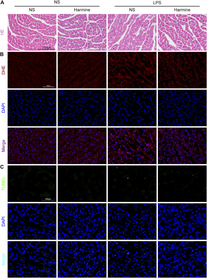
FIGURE 5.
Harmine restrained sepsis-induced cardiac disarrangement, oxidative stress and apoptosis. (A) Representative images of H&E staining in the myocardium (400X, scale bar: 100 μm). (B) Representative images of DHE staining (200X, scale bar: 200 μm). DHE (red) and DAPI (blue). (C) Representative images of TUNEL staining in the myocardium (200X, scale bar: 100 μm). TUNEL (green) and DAPI (blue).

