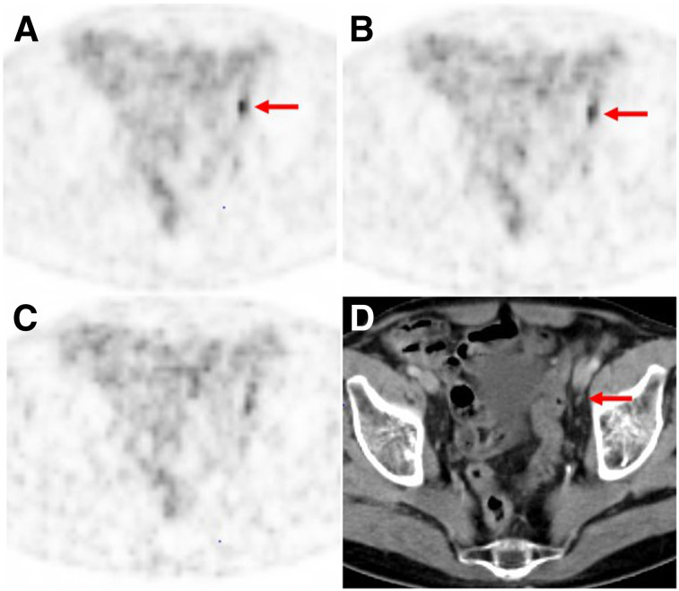
FIGURE 3.
68Ga-PSMA-11 PET/CT images of 75-y-old patient (patient 7) with biochemical recurrence after radical prostatectomy (PSA value, 4 ng/mL at time of imaging). (A and B) Lymph node metastasis (arrows) was classified correctly by all 3 readers on PET performed with standard dose (A) and low dose (B), although mean lesion detectability decreased from 2.67 for standard dose to 2.33 for low dose. (C) On PET performed with very low dose, 2 readers missed this lesion because of increased image noise (mean lesion detectability, 0.67). (D) Corresponding CT image shows small, morphologically unobtrusive lymph node (arrow).

