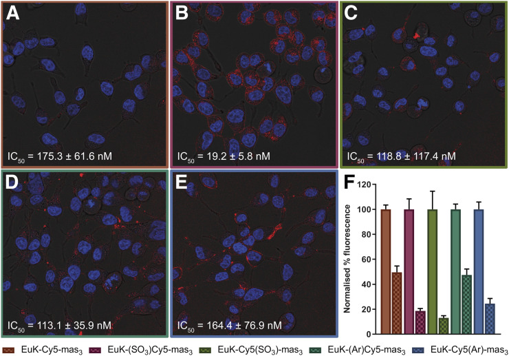
FIGURE 2.
In vitro assessment of hybrid tracers. Shown is staining of extracellular expression of PSMA on C2-2B4 cells after incubation with nuclear stain (Hoechst; in blue) and hybrid tracer (Cy5; in red), with half-maximal inhibitory concentrations (IC50, measured with 125I-EuK–I-BA [((S)-1-carboxy-5-(4-(iodo-benzamido)pentyl)carbamoyl)-l-glutamic acid] as competitive radioligand). (A) EuK-Cy5-mas3. (B) EuK-(SO3)Cy5-mas3. (C) EuK-Cy5(SO3)-mas3. (D) EuK-(Ar)Cy5-mas3. (E) EuK-Cy5(Ar)-mas3. (F) Blocking (checkered) of PSMA with EuK–I-BA. Binding affinities are represented as mean ± SD.

