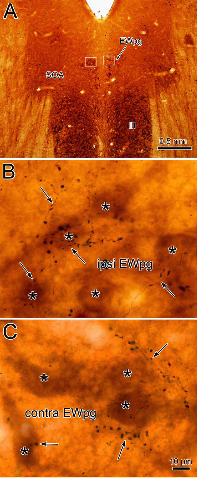
Figure 11.

TLC terminals in the EWpg. (A) Cytochrome oxidase–stained sections reveal the location of the III and the EWpg. Boxes indicate the regions shown in B and C. Examples of the BDA-labeled axonal arbors (arrows) observed in the EWpg in the side ipsilateral (B) and contralateral (C) to the injection site (illustrated in Fig. 10). Large numbers of boutons are present in the vicinity of cytochrome oxidase–labeled presumed preganglionic motoneurons (asterisks). Scale in B = C.
