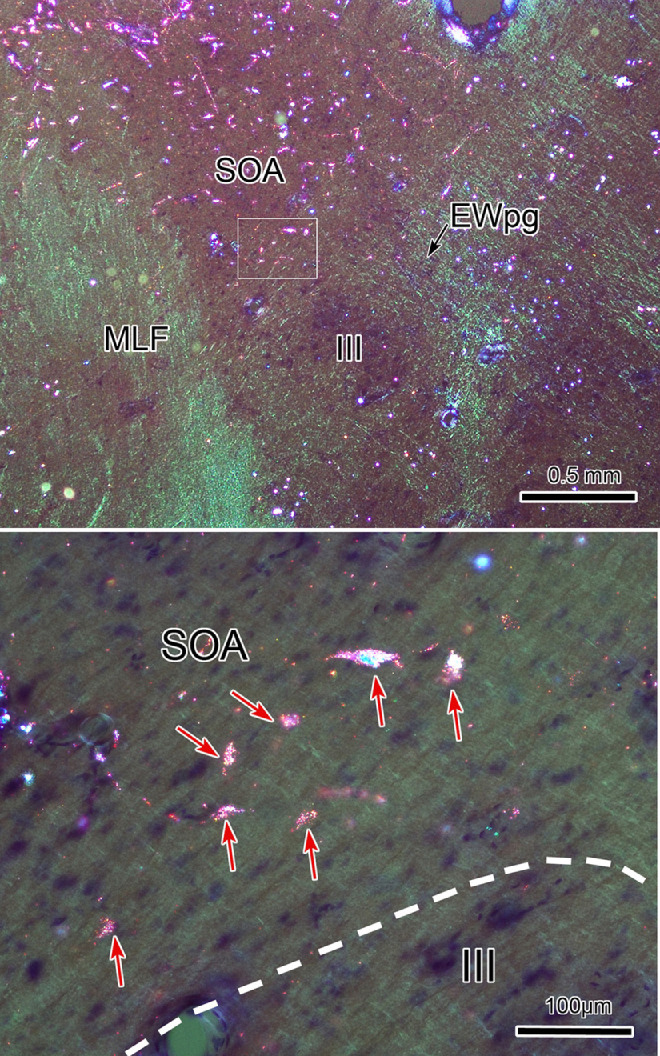
Figure 13.

MPt-TLC afferent neurons in the SOA. Following the WGA-HRP injection shown in Figure 12, retrogradely labeled neurons (B, arrows) were numerous in the SOA. A shows the region containing SOA indicated by a box in Figure 12D. The boxed area in A is presented at a higher magnification in B. Retrograde tracer is revealed by crossed polarizer illumination.
