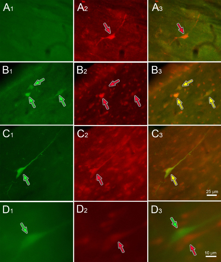
Figure 14.
Some TLC neurons project to the Edinger–Westphal nuclei on both sides. Recombinant rabies virus injections were made into each ciliary body, with N2c-GFP injected on the left and Ns2c-mCherry injected on the right. Fluorescent images showing the GFP (left column), mCherry (middle column), and merged image (right column) are displayed. Green arrows indicate GFP-labeled neurons, and red arrows indicate mCherry-labeled neurons. Both singly labeled (A, D) and doubly labeled (yellow arrows) (B, C) were present. Scale in C3 = A1–3, B1–3, C1–2; in D3 = D1–2.

