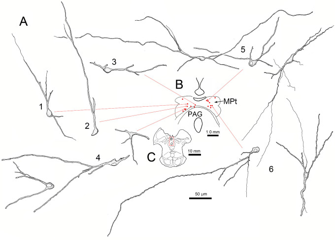
Figure 5.
Morphology of lens premotor neurons in the MPt. Same case as shown in Figure 4. Illustrations show examples of the somata and the dendritic arrangements of rabies-positive neurons (A1–6) present in a single section (B, red dots) through the MPt. (C) Section containing labeled cells has red square to indicate sampled area in B.

