
Figure 9.
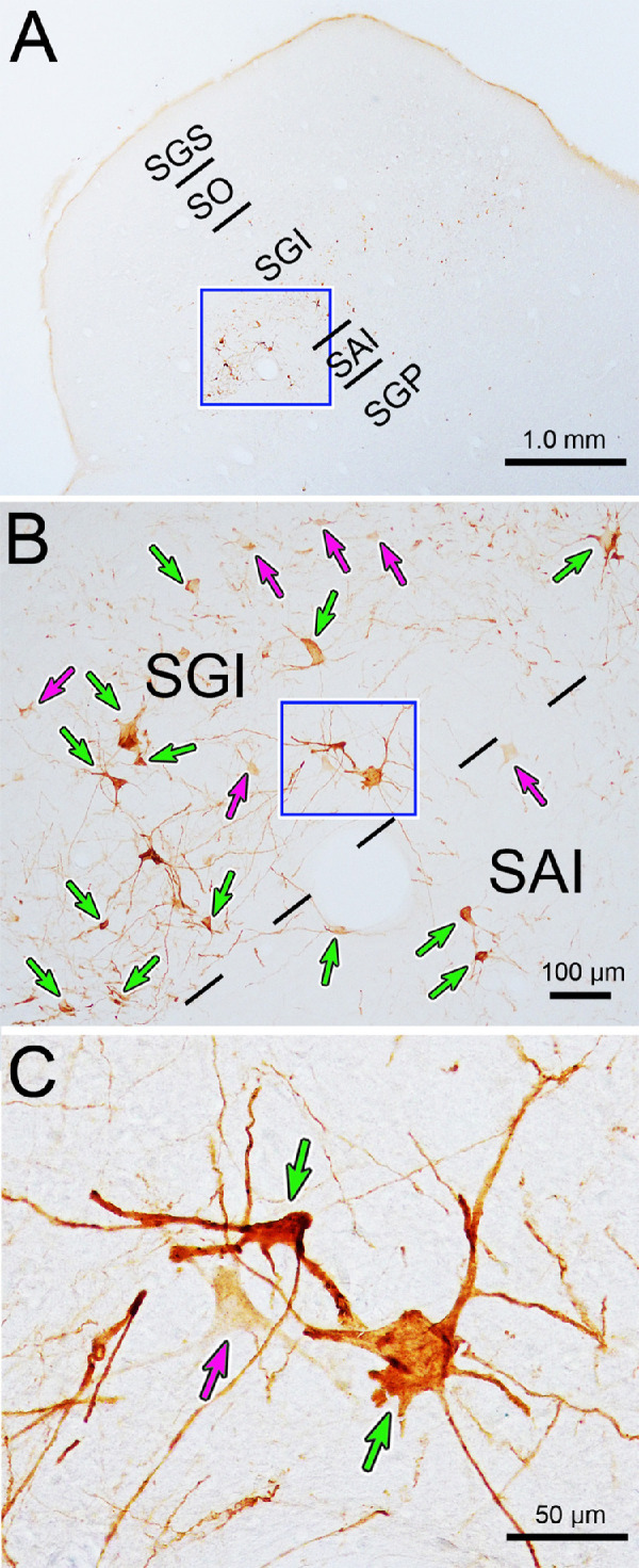
Somatodendritic morphology of neurons labeled in the intermediate gray layer (SGI) 84 hours after a rabies injection of the ciliary body. (A) Low-magnification view showing location of labeled neurons, primarily in the intermediate gray layer (SGI). Box indicates area sampled in B. Both densely labeled (green arrows) and lightly labeled (magenta arrows) neurons are present in the SGI and the intermediate white layer (SAI), as shown in B. Box indicates area sampled in C. The labeled neurons have multipolar somata, with numerous sparsely branched dendrites, as evident in C.