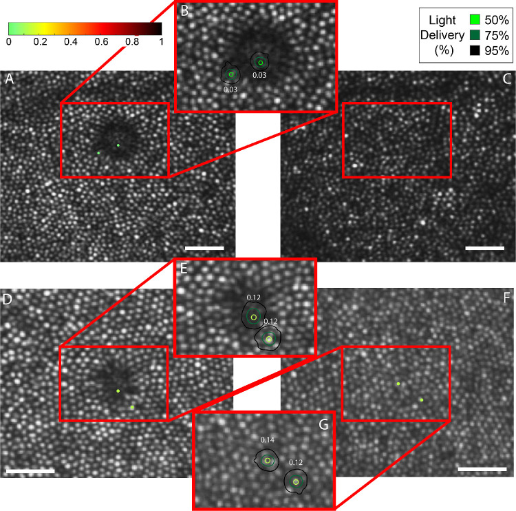
Figure 5.
Microperimetry results for subject 20190 (top) and 20114 (bottom). The tested locations are indicated by dots in the larger field image and by contours in each magnified inset where the color of the dot and the central contour depicts the threshold as indicated by the color bar. Panels (C) and (F) to the right show the follow-up AOSLO images (3 weeks for 20190 and 2 weeks for 20114) after the first microperimetry session where the dysflective cones have recovered their reflectivity. Here, the red box indicates the same location as the red box in (A) and (D). (F) also shows the microperimetry results in the location with recovered reflectivity. The scale bars in each image are 0.1°.

