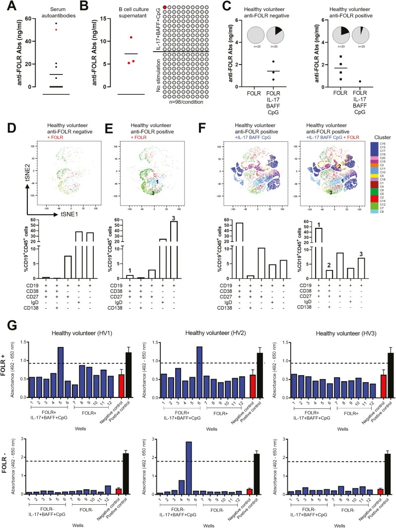
Fig. 4.
Tumour cell- and antigen-reactive antibodies can be detected in the supernatants of IL-17+BAFF+CpG-stimulated and antigen-stimulated B cells. (A) FOLR ELISA assays of serum samples from healthy volunteers (n = 15) show detectable autoantibodies against the tumour-associated antigen folate receptor alpha (FOLR) in five individuals. (B) FOLR ELISA assays of B-cell culture supernatants from a healthy volunteer with detectable serum anti-FOLR autoantibodies (red in A). In three independent experiments performed from the B-cell cultures of the same healthy volunteer, we tested n = 98 wells per independent experiment. This confirmed secretion of FOLR-specific IgG from ex vivo cultured B cells stimulated with IL-17+BAFF+CpG (n = 98 wells), but not from unstimulated B-cell cultures (n = 98 wells) (left: positive well confirmed in all three independent experiments performed from the same healthy volunteer; right: schematic of n = 98 wells per experiment). (C) B cells cultured with human recombinant antigen FOLR in the presence or absence of IL-17+BAFF+CpG led to anti-FOLR-specific antibodies in healthy volunteers with and without previously detectable serum autoantibodies (n = 2; 20 samples/condition, pie charts show proportion of positive cultures [black]). (D–E) Immunophenotyping (tSNE [top] and B-cell subset [bottom]) analysis of B cells from a healthy volunteer with no detectable serum anti-FOLR Abs (D), and from an individual with detectable serum anti-FOLR Abs (E), following ex vivo B-cell stimulation with recombinant FOLR. (F) Immunophenotyping (tSNE [top] and B-cell subset [bottom]) analysis of B-cell populations in a healthy volunteer following activation with IL-17+BAFF+CpG in the absence (left) or presence (right) of FOLR. (G) B cells cultured with IL-17+BAFF+CpG and the human recombinant antigen (FOLR) revealed secreted IgG antibodies binding to FOLR-expressing IGROV1 tumour cells. IGROV1 cell-based ELISA for the assessment of IGROV1-reactive IgG antibodies from culture supernatants of B cells from three healthy volunteers. B cells were stimulated with IL-17+BAFF+CpG and FOLR (top panel) or without FOLR (bottom panel). Assays were performed from culture supernatants collected on day 7. Each blue bar chart represents a supernatant sample from one B-cell culture well (blue). Controls (n = 3; mean ± SEM): Positive control anti-FOLR IgG, MOv18 clone, 400 ng/ml (black); Negative control, non-specific human IgG, 400 ng/ml (red). Threshold for IGROV1 reactivity was set at 75% the optical density (OD) value obtained for the positive control.

