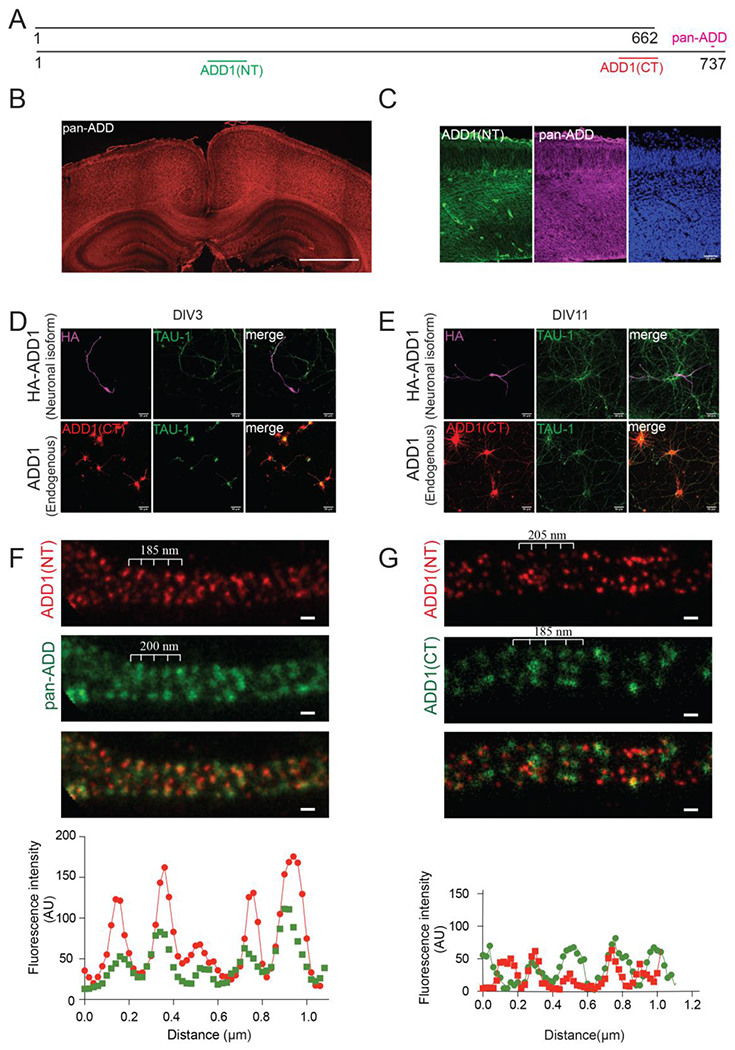
Figure 3. Expression of ADD1 splice isoforms in the brain.

A) Schematic structure of ADD1 isoforms with regions that are recognized by different antibodies. ADD1(NT), sc33633; ADD1(CT), HPA035873; pan-ADD, ab51130.
B) Immunostaining of P7 mouse brain with pan-ADD antibody showing that adducins are expressed in the cortex and enriched in the corpus callosum. Scale bar 1000μm.
C) Immunostaining of E14.5 mouse cortex with pan-ADD and Add1-specific antibody (ADD1(NT)) showing that Add1 is expressed in the developing mouse brain.
D) Immunostaining of transfected and endogenous Add1 showing localization in axons of DIV3 primary mouse hippocampal neurons.
E) Immunostaining of Add1 showing localization in axons of rat DIV11 primary hippocampal neurons.
F) STED imaging of Add1 isoforms in primary cultured rat hippocampal neurons showing periodic signals of Add1(ADD1(NT)) and pan-adducins (pan-ADD) in the axon. Scale bar 0.2 μm.
G) STED imaging of Add1 isoforms in primary cultured rat hippocampal neurons showing periodic signals of Add1 (ADD1(NT), ADD1(CT)) in the axon. Scale bar 0.2 μm.
See also Figure S3.
