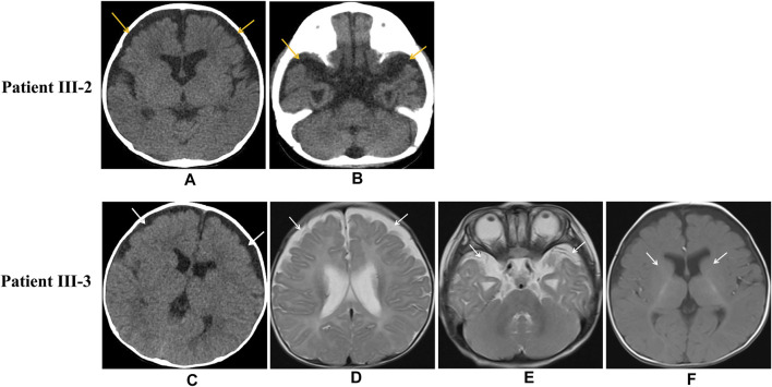
FIGURE 2.
Imaging of patient III-2 and patient III-3. Unenhanced head CT indicates widening of the bifrontal frontotemporal subarachnoid space in patient III-2 (Figures 2A,B) Unenhanced head CT of patient III-3 shows bifrontal widening frontotemporal of the subarachnoid space (Figure 2C), and axial MRI shows bifrontal widening frontotemporal of the subarachnoid space (Figures 2D,E) and delayed myelination of white matter (Figure 2F).

