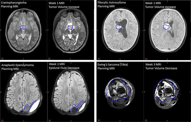
Figure 2.
Planning CT and MR images of an 11-year-old patient with infratentorial ependymoma. 3D T1 TFE: TR/TE = 8/3.5 ms, 1 average, flip angle = 8°, 2-mm slice thickness. 3D T2 TSE: TR/TE = 2500/229 ms, flip angle = 90°, 3 averages, 2-mm slice thickness. 3D T2 FLAIR with fat saturation: TR/TE = 4800/316 ms, 2 averages, 2-mm slice thickness. Diagnostic 3D T1 MPRAGE: TR/TE/TI = 1800/2.26/900 ms, flip angle = 9°, 1 average, 1-mm slice thickness. The yellow contour on CT represents the gross tumor volume.

