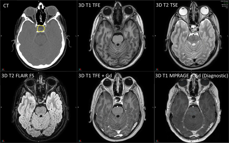
Figure 5.
Planning CT and MR images of a 15-year-old patient with rhabdomyosarcoma. 3D T1 FFE with fat saturation: TR/TE = 4.5/2.2 ms, flip angle = 10°, 4 averages, 2.4-mm slice thickness. 3D T2 TSE with fat saturation: TR/TE = 2000/191 ms, flip angle = 90°, 2 averages, 2.4-mm slice thickness. Diagnostic axial T1 FLAIR with/without fat saturation: TR/TE/TI = 3250/9.4/1000 ms, flip angle = 140°, 1 average, 3-mm slice thickness. Diagnostic axial T2 TSE with fat saturation: TR/TE = 4930/93 ms, flip angle = 150°, 2 averages, 3-mm slice thickness.

