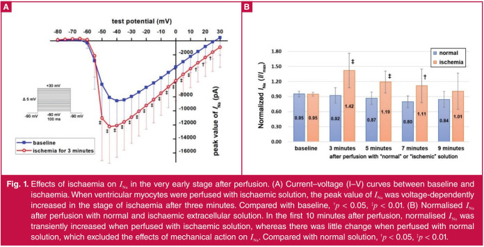
Fig. 1.
Effects of ischaemia on INa in the very early stage after perfusion. (A) Current–voltage (I–V) curves between baseline and ischaemia. When ventricular myocytes were perfused with ischaemic solution, the peak value of INa was voltage-dependently increased in the stage of ischaemia after three minutes. Compared with baseline, †p < 0.05, ‡p < 0.01. (B) Normalised INa after perfusion with normal and ischaemic extracellular solution. In the first 10 minutes after perfusion, normalised INa was transiently increased when perfused with ischaemic solution, whereas there was little change when perfused with normal solution, which excluded the effects of mechanical action on INa. Compared with normal solution, †p < 0.05, ‡p < 0.01.

