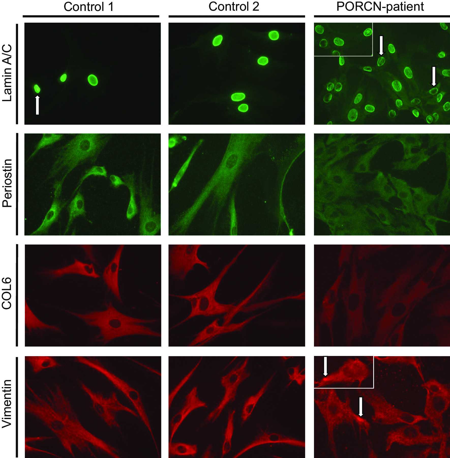
Fig. 4.

Immunological studies to confirm proteomic data: Immunofluorescence studies revealed the presence of irregular nucleoplasmic Lamin A/C-depositions (often appearing as rods) in cells derived from the patient in addition to nuclei with remarkable decreased immunoreactivity (white arrows). Periostin and Collagen alpha-3 (VI) decreased cellular level in patient-derived cells. Vimentin staining revealed a uniform distribution throughout the cytoplasm of control fibroblasts, in patient-derived cells, the fluorescence intensity of reticular Vimentin staining is less intense. Moreover, focal increase of Vimentin within the cytoplasm and adjacent to the plasma membrane was frequently identified in patient-derived fibroblasts. Scale bars = 30 µm
