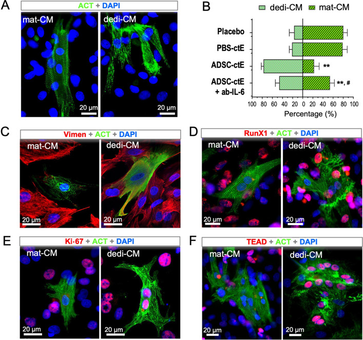
Fig. 5.
Obligatory role of IL-6 in the induction of cardiac dedifferentiation in isolated neonatal cardiomyocytes. A In cultured neonatal cardiomyocytes, existence of mature cardiomyocytes (mat-CM) and dedifferentiated cells (dedi-CM) was categorized by cardiac a-actinin (ACT) staining upon their distinct morphological appearance: the mat-CM showed a clear striation pattern, while the dedi-CM featured a sarcomeric assembly. B Spontaneous dedifferentiation in the cultured neonatal cardiomyocytes after 5-day cultivation was estimated at the rate of 20% in placebo controls (n = 5). Addition of tissue extracts from PBS control hearts (PBS-ctE) did not alter the dedifferential rate, but when cardiac tissue extracts from ADSCgfp+-treated hearts (ADSC-ctE, 10%, n = 5), was supplemented, the rate was accelerated up to 78%, leading to the ratio of both types of CM significantly shifted to dedi-CM dominating statue in the culture. The proportional shift of two populations was partially reverted by supplement of monoclonal antibody against IL-6 (ab-IL-6, n = 4). C–F While the mat-CM showed almost negative staining for mesenchymal cells (Vimen and RunX1), cell proliferation (Ki-67) and YAP-responsive target (TEAD1), the dedi-CM cells were stained positive for those markers, which was found likewise in vivo situations. **p < 0.01 compared to the PBS-ctE treated cells and #p < 0.01 compared to ADSC-ctE treated cells

