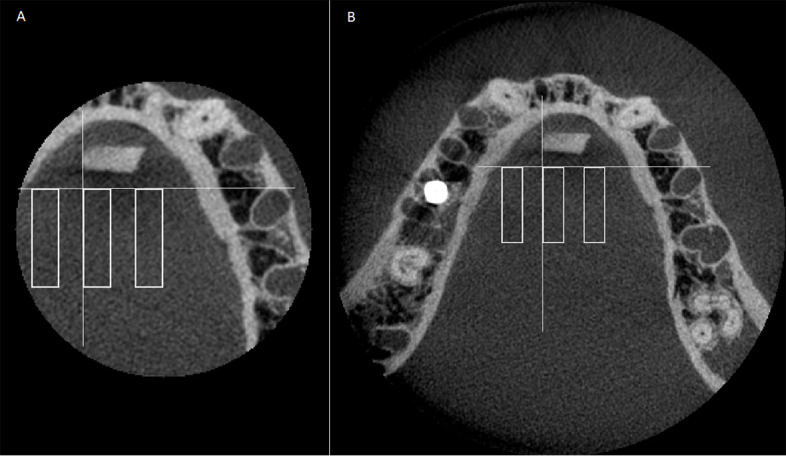
Figure 3.

CBCT axial images indicating the three rectangular regions of interest: A. 4 × 5 cm FOV; B. 8 × 5 cm FOV. White lines are references for the placement of the middle ROI. ROI were established close to the implant, in the middle, and further from the implant, using a line tangent to the alveolar socket of second left premolar and a line perpendicular to the first and tangent to the ERBS block. From the intersection of those lines, the middle ROI was established. The close and further ROIs were drawn 3.6 mm from the middle ROI. . CBCT, cone-beam CT; FOV, field of view; ROI, region of interest.
