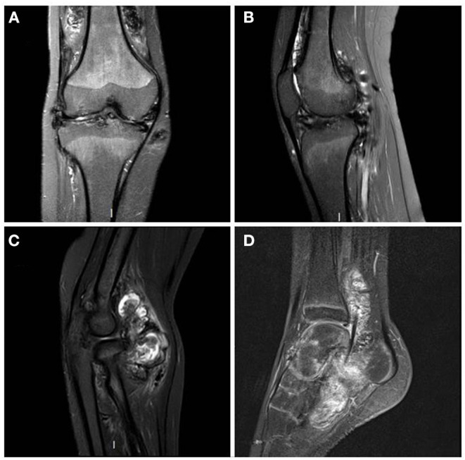
Figure 1.

MRI T1 Fat Sat sequences - right knee in coronal plane (A) and right knee in sagittal plane (B): diffuse synovial pannus, especially in the superior recess of the joint. Tri-compartmental chondropathy associating stage IV chondrolysis associated with multiple subchondral cysts; STIR sequence of the right elbow in sagittal plane (C): anterior intra-articular synovial masses in heterogeneous signal; T1 sequence after gadolinium injection of left ankle in sagittal plane (D): Large synovial panuses mainly affecting the subtalar joint and voluminous synovial masses developed around the long plantar flexor hallucis behind the lower end of the tibia. These signs were highly suggestive of multifocal villonodular synovitis which was later confirmed by histopathological examination.
