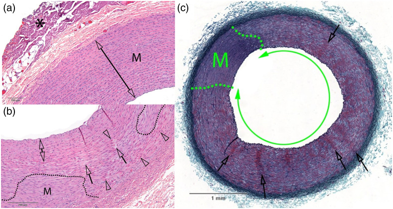
Fig. 3.
Examples of carotid artery vessels subjected to various dosages of light radiation after staining with (a), (b) H&E and (c) GET. The dosages by panel are (a) 1720 nm, ; (b) 1720 nm, ; and (c) 1720 nm, . The dotted lines separate intact regions (M) of vessel tissue from damaged regions. The asterisk in (a) marks collagen denaturation, likely from electrocauterization used to remove tissue during the surgery. Otherwise, no damage was detected in (a). In (b), clear arrowheads indicate hypereosinophilic and contracted SMCs, whereas clear arrows show regions with cell effacement. The double arrow indicates a region with pyknotic nuclei and hypereosinophilic SMCs, consistent with compressive injury. Damage is present throughout the vessel in (c), in the form of widespread media necrosis (double green arrow). In addition, clear arrows show radial clusters of hyperchromatic and shrunken SMCs evoking contraction bands. Scale bar shows physical size of carotid artery. Additional notated images of samples subjected to these dosages are shown in the pathology report in the Supplementary Material.

