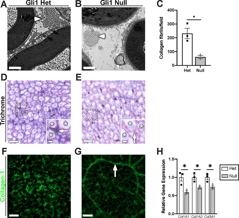
Figure 9.
Gli1 expression controls peripheral nerve ECM. A, B, High-magnification EMs reveal a decrease in collagen fibrils (arrowheads) in the Gli1 nulls versus hets. C, Quantification of collagen fibril density demonstrates a significant decrease in Gli1 nulls (60.67 ± 11.05) compared with hets (231.7 ± 37.55) (p = 0.036). D, E, Gli1 het and null sciatic nerves were stained with Masson's trichrome, which labels collagen blue. Staining is decreased in the extracellular spaces (insets, arrows) between axons (insets, stars) of Gli1 nulls. F, G, Gli1 het and null nerves were stained with an antibody against full-length (native) collagen-1 (green). Staining intensity is decreased in the endoneurium of Gli1 nulls, although MFs are moderately stained (arrow). H, Transcript levels for several key collagen genes were determined by qPCR and show significant decreases for the three main fibril-forming collagen genes expressed in the endoneurium. COL1A1: Het (1.00 ± 0.10) versus Null (0.59 ± 0.05) (p = 0.042); COL2A1: Het (1.00 ± 0.07) versus Null (0.72 ± 0.033) (p = 0.035); COL3A1: Het (1.00 ± 0.06) versus Null (0.74 ± 0.05) (p = 0.033). Values shown represent mean ± SEM analyzed using unpaired t test with Welch's correction based on n = 10 fields per replicate (C) and n = 3 biological replicates per genotype (C,H). *p < 0.05. Scale bars: A, B, 0.5 μm; D, E, 10 μm; D, E, insets, 2 μm; F, G, 10 μm.

