Figure 3.
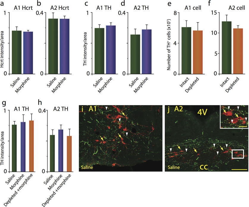
Morphine treatment does not alter Hcrt axon density, TH levels, or TH cell numbers in the A1/A2 medullary norepinephrine-containing regions. a, b, Intact-DTA-Hcrt animals treated with morphine (14 d, 50 mg/kg) did not show a significant change in Hcrt axonal labeling in the A1 (a) and the A2 (b) regions. c, d, TH levels remained comparable to baseline levels after morphine treatment in the A1 (c) and A2 (d) regions. e, f, TH+ cell counts were unaffected by the elimination of Hcrt neurons in depleted-DTA-Hcrt animals in the A1 (e) or the A2 (f) regions. g, h, Deletion of Hcrt neurons did not affect TH levels in the A1 (g) and the A2 (h) regions after morphine treatment compared with intact-DTA-Hcrt animals treated with saline or morphine. i, j, Confocal images of representative sections of the A1 (i) and A2 (j) of an intact-DTA-Hcrt animal after saline treatment, illustrating the distribution of Hcrt fibers in green (white arrowheads) and TH+ neurons in red (yellow arrows). Insert in j shows a higher magnification of white square. Scale bars: 100 µm; insert, 20 µm. cc, Central canal; 4V, fourth ventricle. n = 4/condition.

