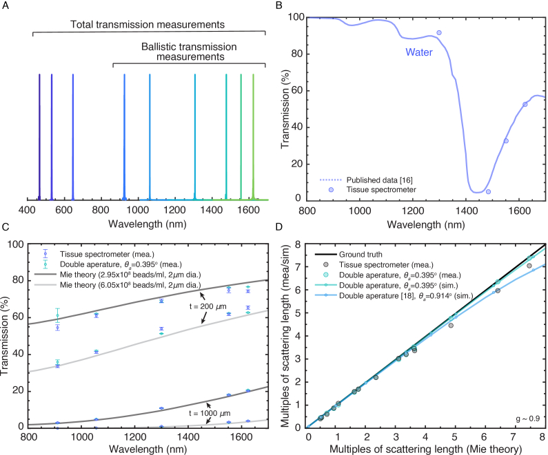
Fig. 2.
Wavelength selection and validation of the tissue spectrometer accuracy. (A) Wavelength measurements of the CW lasers used. (B) Total transmission of water measured by the tissue spectrometer (circle dot) compared to previous measurements [16]. (C) Ballistic transmission of four tissue phantoms measured by the tissue spectrometer (purple error bar) and compared to the Mie theory (solid line) and measurements using a double-aperture setup at 0.395° acceptance angle (green error bar). (D) Comparison of the retrieved multiples of scattering length to the predicted multiples of scattering length. Mea. = measurement, sim. = simulation.

