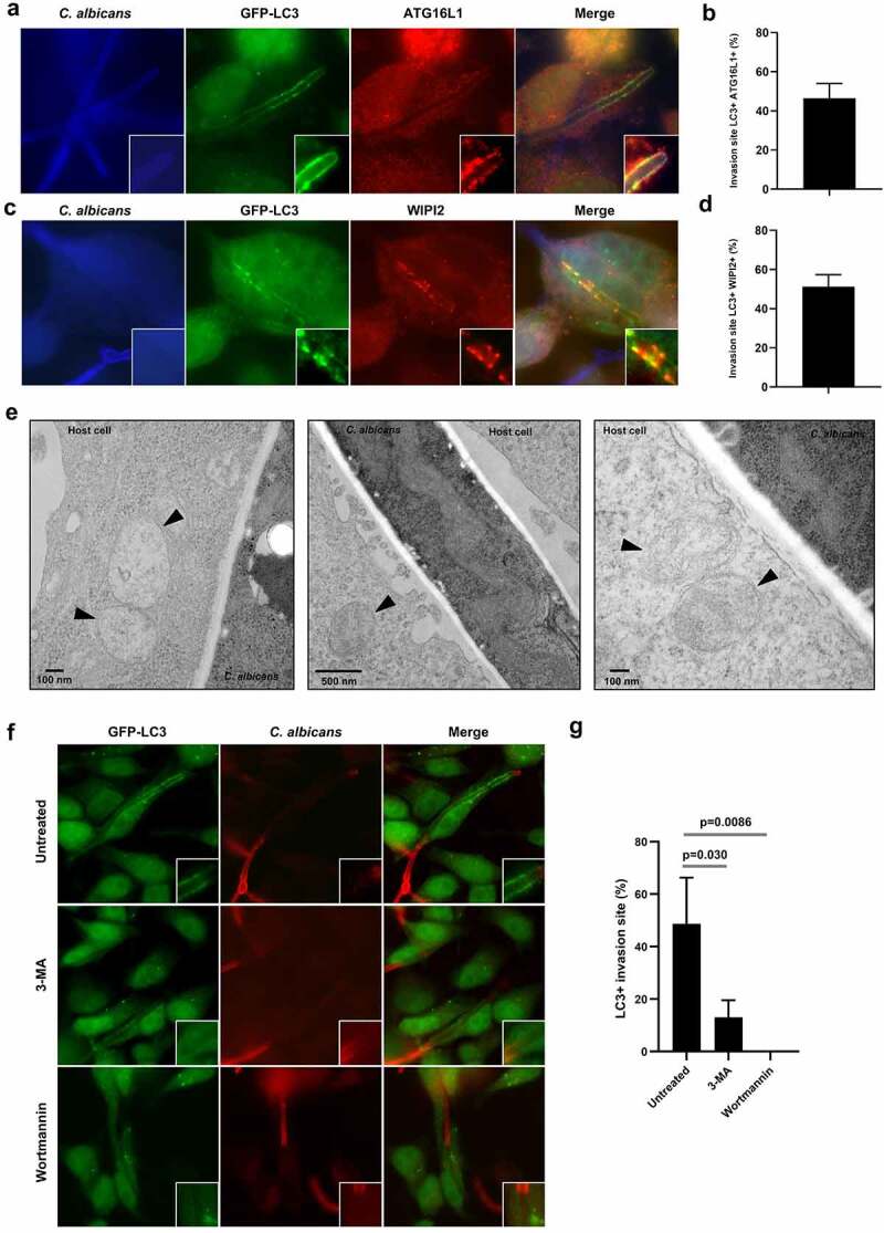
Figure 2.

Key components of the autophagy machinery are mobilized in the vicinity of the C. albicans entry sites.
(a and c) Representative images of C. albicans-infected GFP-LC3 HeLa cells after 4 h infection. Samples were processed for C. albicans (blue), GFP-LC3 (green), and (a) ATG16L1 (red) or (c) WIPI2 (red) staining. (b and d) Quantification of the percentage of (b) GFP-LC3+/ATG16L1+ or (d) GFP-LC3+/WIPI2 + C. albicans invasion sites at 4 h postinfection. Data are mean ± SEM of three independent experiments. (e) Ultrastructural analysis by transmission electron microscopy of HeLa cells infected with C. albicans for 4 h showing the presence of intracellular vacuoles with autophagosome features (indicated by black arrowheads) near Candida invasion sites. (f) Representative images of GFP-LC3 HeLa cells untreated or treated with the PI3K inhibitors 3-MA (5 mM) or Wortmannin (100 nM) and infected with C. albicans for 4 h. Samples were processed for C. albicans (red) and GFP-LC3 (green) staining. (g) Quantification of the percentage of GFP-LC3 + C. albicans invasion sites at 4 h postinfection. Data are mean ± SEM of three independent experiments.
