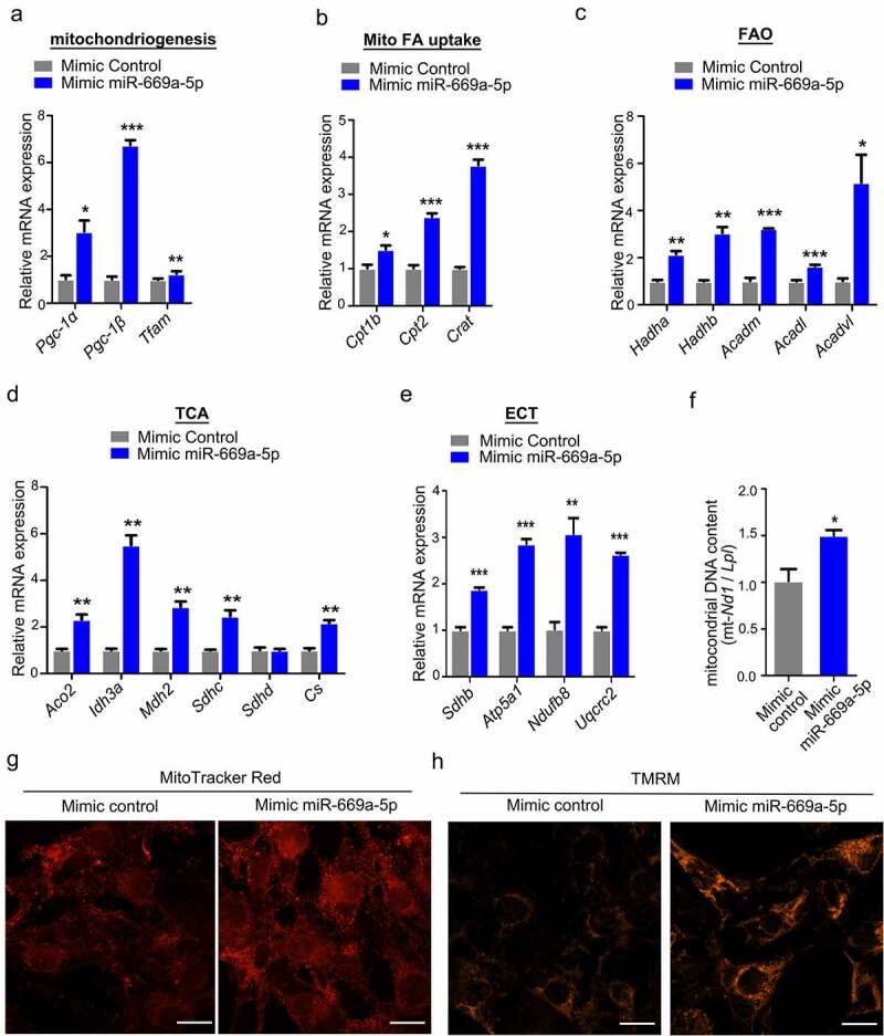
Figure 6.

miR-669a-5p enhances the mitochondrial biogenesis in 3T3-L1 cells. 3T3-L1 preadipocytes were transfected with mimic control or mimic miR-669a-5p (100 nM) on day 0 and day 4 after differentiation, the cells were collected on day 8 for analysis.(a) RT-qPCR analysis of mitochondrial genes involved in mitochondriogenesis, normalized to β-actin expression. n = 3 per group.(b) RT-qPCR analysis of mitochondrial genes involved in fatty acid uptake (Mito FA uptake) in mature adipocytes, normalized to β-actin expression. n = 3 per group.(c) RT-qPCR analysis of mitochondrial genes involved in β-oxidation (FAO) in mature adipocytes, normalized to β-actin expression. n = 3 per group.(d) RT-qPCR analysis of representative tricarboxylic acid (TCA) cycle genes in mature adipocytes, normalized to β-actin expression. n = 3 per group.(e) RT-qPCR analysis of representative genes of the five complexes of the mitochondrial electron transport chain (ETC) in mature adipocytes, normalized to β-actin expression. n = 3 per group.(f) The quantification of mitochondrial DNA content. n = 3 per group(g) Mitochondrial mass was measured by MitoTracker Red staining on day 8. n = 3 per group.(h) Mitochondrial membrane potential was measured by TMRM on day 8. n = 3 per group.Data are representative of at least three individual experiments. Results are represented as mean ± SEM. *p < 0.05, **p < 0.01, ***p < 0.001 versus mimic control group. Scale bar indicates 20 μm in G and H.
