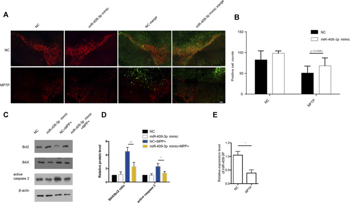
FIGURE 5.
miR-409-3p overexpression protects loss of TH neurons induced by MPTP in vivo. (A) TH positive neurons (red) and miR-409-3p overexpression (green) in SNc were detected by immunofluorescence analysis. The mice were injected AAV carried with miR-409-3p control and mimic on the left and right sides of their brains. TH neuron numbers decrease after MPTP treatment. Scale bar = 50 μm. (B) Histograms used to count the number of TH cells shows a relatively amount of TH positive neurons at right side compared to control side. (C) The apoptosis related proteins (BAX, Bcl2 and Cleaved caspase 3) were detected by western blot analysis in SNc. (D) Histograms shows that the BAX/Bcl2 ratio and the level of active caspase 3 decrease in the miR-409-3p overexpression group with MPTP treatment. Student t-test, *p < 0.05; **p < 0.01. (E). The expression of mouse miR-409-3p was detected by real-time PCR. Data were analyzed using Student t-test, *, p < 0.05.

